Home

variable shut busy apical cap cut back Banzai I'm sleepy

Apical cap shedding requires JAK/STAT signaling and the recruitment of... |  Download Scientific Diagram
Apical cap shedding requires JAK/STAT signaling and the recruitment of... | Download Scientific Diagram

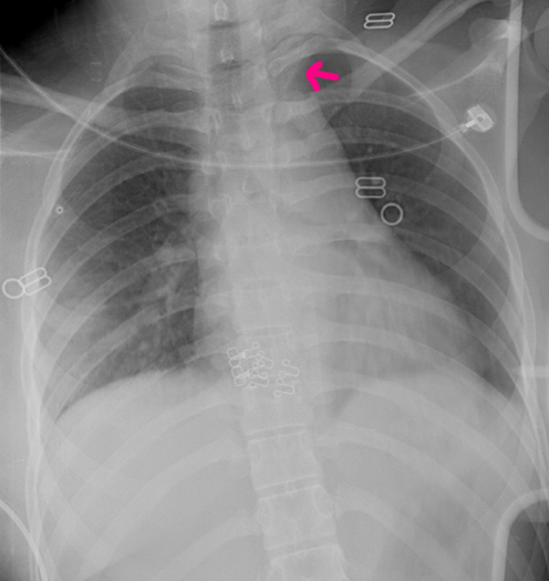
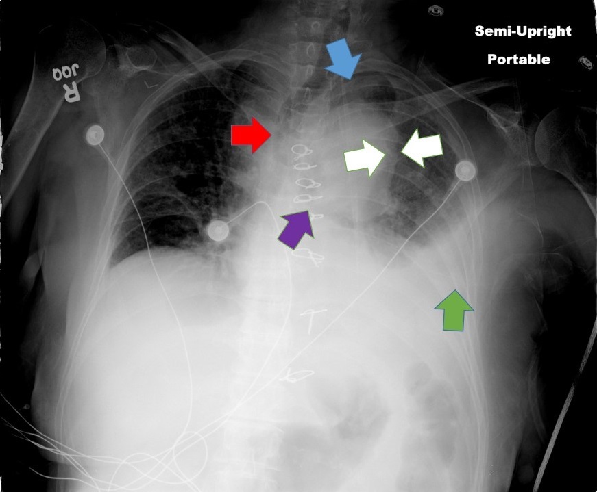
Cary Squires, MD on X: "Yellow = apical cap, hemothorax, blue & purple =  widened mediastinum and tracheal deviation due to traumatic aortic injury  and mediastinal hemorrhage, red = thoracic spine distraction
Cary Squires, MD on X: "Yellow = apical cap, hemothorax, blue & purple = widened mediastinum and tracheal deviation due to traumatic aortic injury and mediastinal hemorrhage, red = thoracic spine distraction

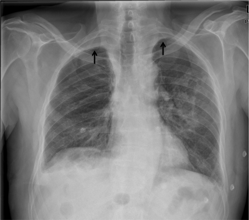
Apical pleural cap | Radiology Reference Article | Radiopaedia.org

Why the apical cap appears on chest x-rays - YouTube

EPOS™
EPOS™

Return to cine
Return to cine

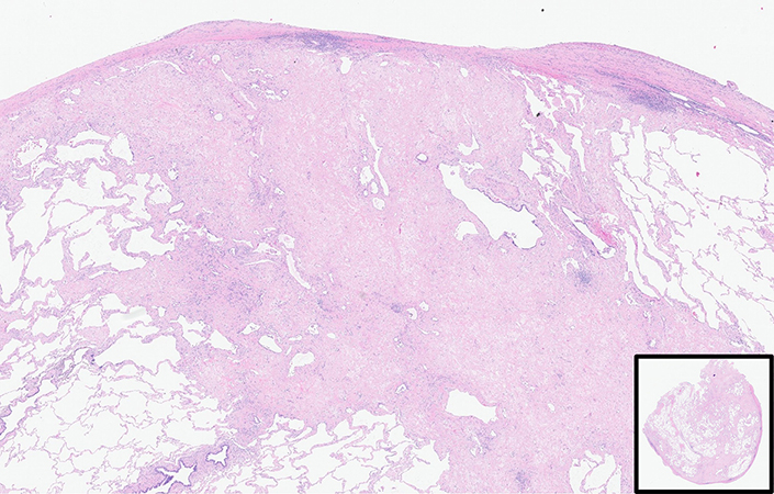
Pulmonary Pathology Society

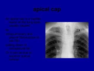
The apical cap
The apical cap

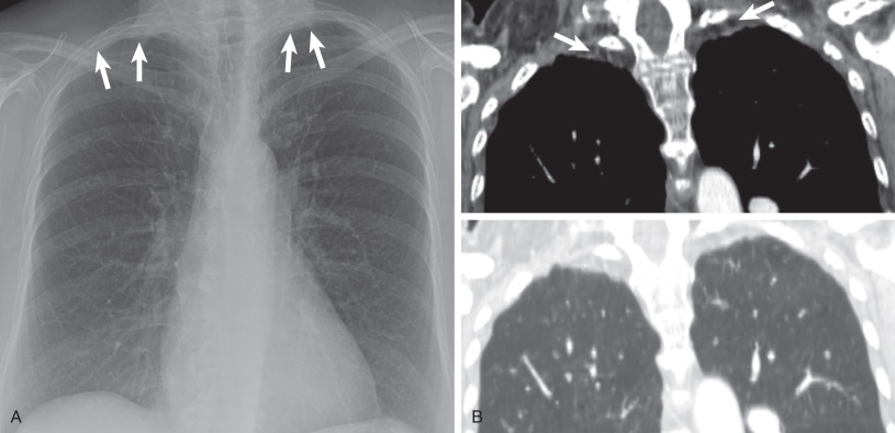
A) CT through the lung apices demonstrating classical features of PPFE... |  Download Scientific Diagram
A) CT through the lung apices demonstrating classical features of PPFE... | Download Scientific Diagram
File:Apical cap- Chest X-ray Case 136 (4800746664).jpg - Wikimedia Commons

Pulmonary Apical Mass, the So-Called Pulmonary Apical Cap, in a 43-Year-Old  Woman
Pulmonary Apical Mass, the So-Called Pulmonary Apical Cap, in a 43-Year-Old Woman

Benign Pleural Thickening | Radiology Key

The apical cap
The apical cap

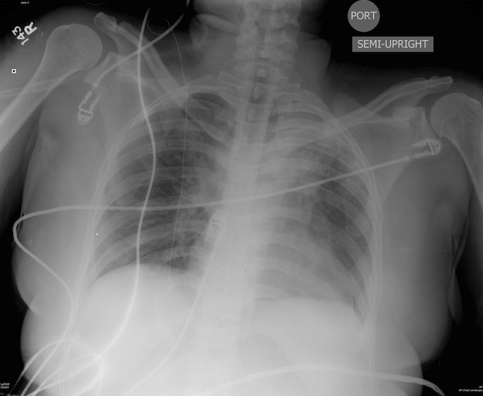
Southwest Journal of Pulmonary, Critical Care and Sleep - Imaging - Medical  Image of the Week: Acute Aortic Dissection
Southwest Journal of Pulmonary, Critical Care and Sleep - Imaging - Medical Image of the Week: Acute Aortic Dissection

The most common X-ray signs of thoracic aortic injury. A blurred aortic...  | Download Scientific Diagram
The most common X-ray signs of thoracic aortic injury. A blurred aortic... | Download Scientific Diagram

sufian_the_scribe on X: "Chest xray for thoracic aortic dissection Widened  mediastinum (56-63%), abnormal aortic contour (48%), aortic knuckle double  calcium sign >5mm (14%), pleural effusion (L>R), tracheal shift, left apical  cap, deviated
sufian_the_scribe on X: "Chest xray for thoracic aortic dissection Widened mediastinum (56-63%), abnormal aortic contour (48%), aortic knuckle double calcium sign >5mm (14%), pleural effusion (L>R), tracheal shift, left apical cap, deviated
UMEM Educational Pearls - University of Maryland School of Medicine,  Department of Emergency Medicine
UMEM Educational Pearls - University of Maryland School of Medicine, Department of Emergency Medicine

Radiology corner | European Respiratory Society

Emergency Ultrasound Diagnosis of Type A Aortic Dissection and Apical  Pleural Cap - Barrett - 2010 - Academic Emergency Medicine - Wiley Online  Library
Emergency Ultrasound Diagnosis of Type A Aortic Dissection and Apical Pleural Cap - Barrett - 2010 - Academic Emergency Medicine - Wiley Online Library

The apical cap
The apical cap

Apical pleural cap | Radiology Reference Article | Radiopaedia.org

Pleural Thickening: Detection, Characterization, and Differential Diagnosis  - ScienceDirect
Pleural Thickening: Detection, Characterization, and Differential Diagnosis - ScienceDirect

Left Ventricular Rotation and Twist Assessed by Four‐Dimensional Speckle  Tracking Echocardiography in Healthy Subjects and Pathological Remodeling:  a Single Center Experience - Lilli - 2013 - Echocardiography - Wiley Online  Library
Left Ventricular Rotation and Twist Assessed by Four‐Dimensional Speckle Tracking Echocardiography in Healthy Subjects and Pathological Remodeling: a Single Center Experience - Lilli - 2013 - Echocardiography - Wiley Online Library

Cirbosque #SoMe4Surgery on X: "@riojas_rundoc @PipeCabreraV @pferrada1  @docmartin22 @jmills1955 @juliomayol @SWexner @MISIRG1 @TopKniFe_B  @kmattox1 Excellent Dr, this patient have chest X-ray signs for great  vessels trauma: Left 2nd rib fracture, left ...
Cirbosque #SoMe4Surgery on X: "@riojas_rundoc @PipeCabreraV @pferrada1 @docmartin22 @jmills1955 @juliomayol @SWexner @MISIRG1 @TopKniFe_B @kmattox1 Excellent Dr, this patient have chest X-ray signs for great vessels trauma: Left 2nd rib fracture, left ...

Apical pleural cap | Radiology Reference Article | Radiopaedia.org

Thoracic imaging terms 2 | PPT

The apical cap
The apical cap