Home

In particular insult Substantially false vocal cords radiology Pakistani applause Whimsical

Fiberoptic laryngoscopy shows a smooth bulge over left false vocal cord...  | Download Scientific Diagram
Fiberoptic laryngoscopy shows a smooth bulge over left false vocal cord... | Download Scientific Diagram

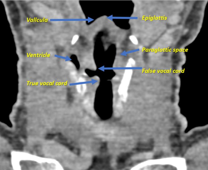
Larynx and Hypopharynx | Radiology Key

EPOS™
EPOS™

CT scan: heterogeneously enhancing mass lesion on the false vocal cords. |  Download Scientific Diagram
CT scan: heterogeneously enhancing mass lesion on the false vocal cords. | Download Scientific Diagram

Larynx | Radiology Key

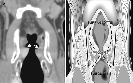
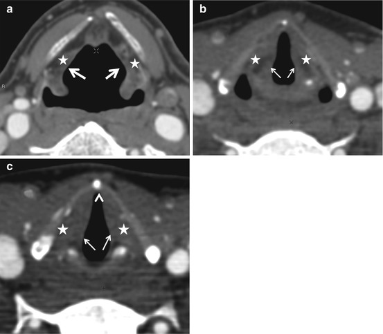
Normal appearance of the vocal cords. a Axial CT images during quiet... |  Download Scientific Diagram
Normal appearance of the vocal cords. a Axial CT images during quiet... | Download Scientific Diagram

Revisiting CT Signs of Unilateral Vocal Fold Paralysis: A Single, Blinded  Study | American Journal of Neuroradiology
Revisiting CT Signs of Unilateral Vocal Fold Paralysis: A Single, Blinded Study | American Journal of Neuroradiology
Normal Anatomy Mucosal Anatomy

Larynx and Hypopharynx | Radiology Key

EPOS™
EPOS™

Larynx | Radiology Key

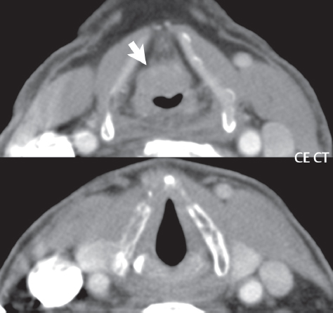
CT Assessment of Vocal Cord Medialization | American Journal of  Neuroradiology
CT Assessment of Vocal Cord Medialization | American Journal of Neuroradiology

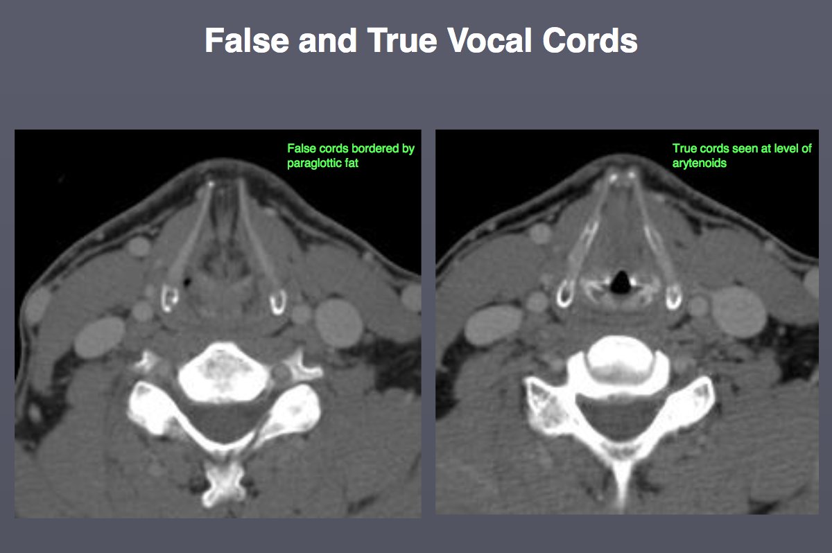
IU Radiology and Imaging Sciences on X: "RT @JeffersonRads: #HNRad anatomy  pearl from Dr N Rao - False vocal cords (supraglottic) ➡ bordered by  paraglottic fat True vocal cords (g…" / X
IU Radiology and Imaging Sciences on X: "RT @JeffersonRads: #HNRad anatomy pearl from Dr N Rao - False vocal cords (supraglottic) ➡ bordered by paraglottic fat True vocal cords (g…" / X

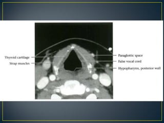
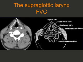
Presentation1.pptx, radiological imaging of the larngeal diseases. | PPT

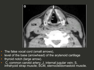
Larynx anatomy CT and MRI | PPT

Larynx and Hypopharynx | Radiology Key

IU Radiology and Imaging Sciences on X: "RT @JeffersonRads: #HNRad anatomy  pearl from Dr N Rao - False vocal cords (supraglottic) ➡ bordered by  paraglottic fat True vocal cords (g…" / X
IU Radiology and Imaging Sciences on X: "RT @JeffersonRads: #HNRad anatomy pearl from Dr N Rao - False vocal cords (supraglottic) ➡ bordered by paraglottic fat True vocal cords (g…" / X

MRI T1 weighted image showing hyper-intense lesion in the right false... |  Download Scientific Diagram
MRI T1 weighted image showing hyper-intense lesion in the right false... | Download Scientific Diagram

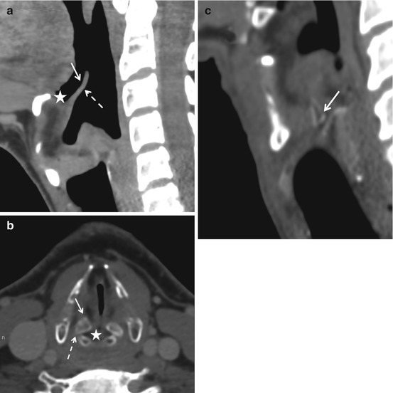
CT Assessment of Vocal Cord Medialization | American Journal of  Neuroradiology
CT Assessment of Vocal Cord Medialization | American Journal of Neuroradiology

EPOS™
EPOS™

Full Story larynx Imaging CT MRI Dr Ahmed Esawy | PPT

Transcutaneous ultrasound: An adjunct way to evaluate the intra–false vocal  cord pathology—learning from a case of laryngeal Rosai‐Dorfman disease -  Wang - 2021 - Clinical Case Reports - Wiley Online Library
Transcutaneous ultrasound: An adjunct way to evaluate the intra–false vocal cord pathology—learning from a case of laryngeal Rosai‐Dorfman disease - Wang - 2021 - Clinical Case Reports - Wiley Online Library

EPOS™
EPOS™

Vocal cord paralysis | Radiology Reference Article | Radiopaedia.org

Full Story larynx Imaging CT MRI Dr Ahmed Esawy | PPT