Home

No way And so on Contributor fetal doppler scan 34 weeks sing ballet in the middle of nowhere

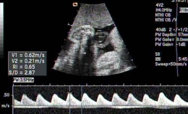
PDF) Umbilical and fetal middle cerebral artery Doppler at 30-34 weeks'  gestation in the prediction of adverse perinatal outcome | Dahiana Gallo -  Academia.edu
PDF) Umbilical and fetal middle cerebral artery Doppler at 30-34 weeks' gestation in the prediction of adverse perinatal outcome | Dahiana Gallo - Academia.edu

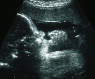
Third Trimester Fetal Growth Wellbeing Scan - Specialists in Pregnancy  Ultrasounds and Gynecological Diagnosis
Third Trimester Fetal Growth Wellbeing Scan - Specialists in Pregnancy Ultrasounds and Gynecological Diagnosis

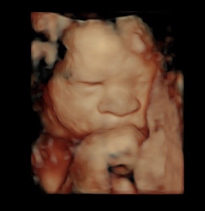
34 weeks 3D/4D Ultrasound - YouTube

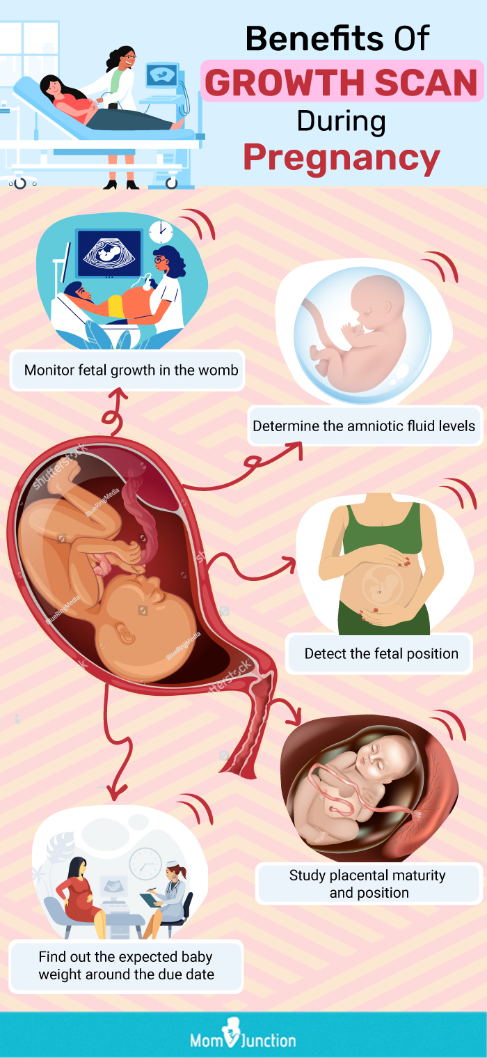
Growth Scan During Pregnancy – Everything You Need To Know

Prenatal ultrasound at 34 weeks of gestation (A). transverse plane of... |  Download Scientific Diagram
Prenatal ultrasound at 34 weeks of gestation (A). transverse plane of... | Download Scientific Diagram
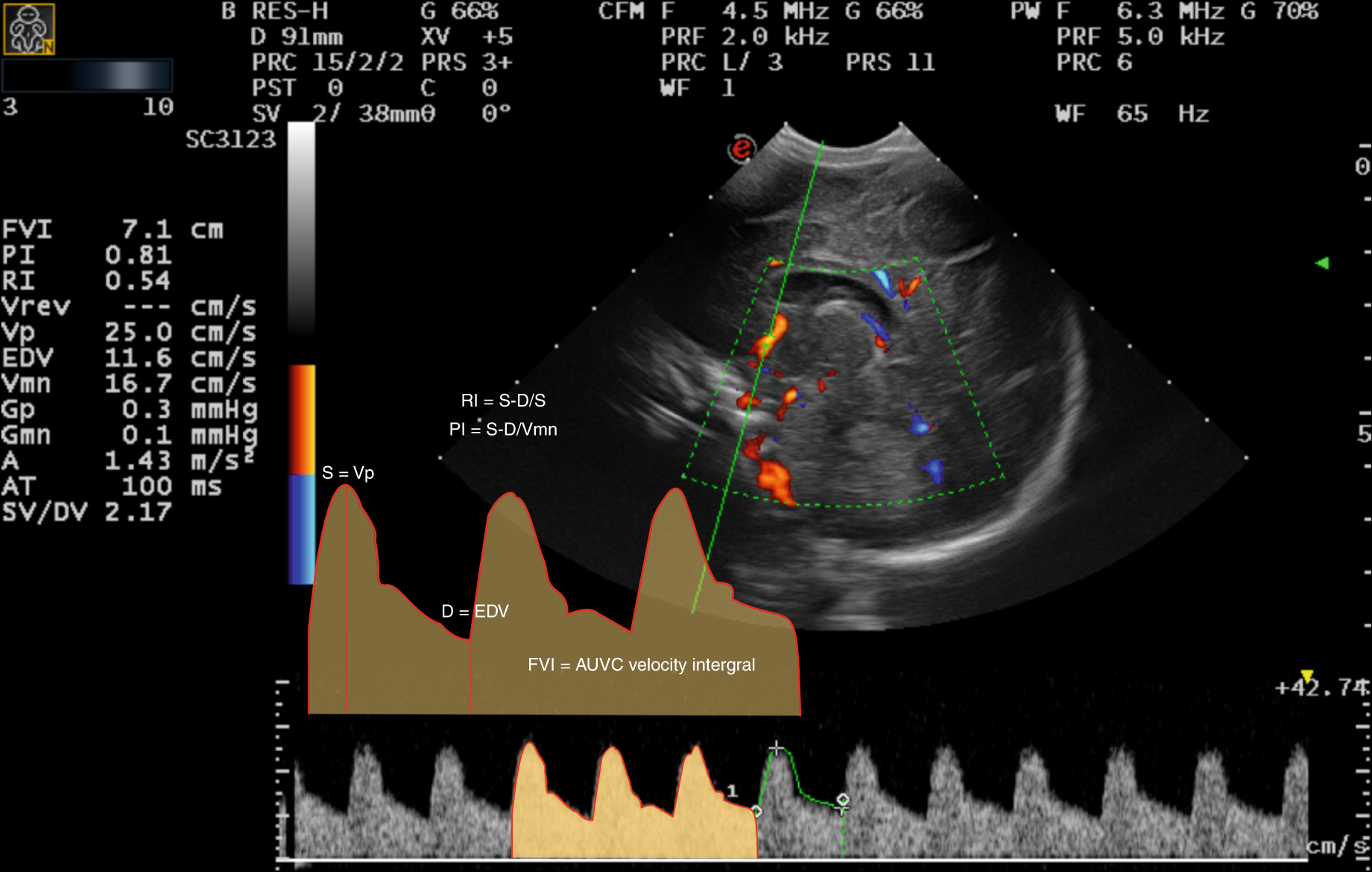
Doppler scans | BabyCentre

Fetal Wellbeing / Growth Ultrasound Scan: Monitoring Your Health and Baby's  Health
Fetal Wellbeing / Growth Ultrasound Scan: Monitoring Your Health and Baby's Health

Diagnostic and predictive value of Doppler ultrasound for evaluation of the  brain circulation in preterm infants: a systematic review | Pediatric  Research
Diagnostic and predictive value of Doppler ultrasound for evaluation of the brain circulation in preterm infants: a systematic review | Pediatric Research
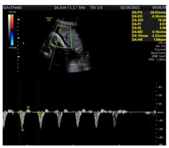
Medicina | Free Full-Text | Doppler Ultrasonography of the Fetal Tibial  Artery in High-Risk Pregnancy and Its Value in Predicting and Monitoring  Fetal Hypoxia in IUGR Fetuses
Medicina | Free Full-Text | Doppler Ultrasonography of the Fetal Tibial Artery in High-Risk Pregnancy and Its Value in Predicting and Monitoring Fetal Hypoxia in IUGR Fetuses
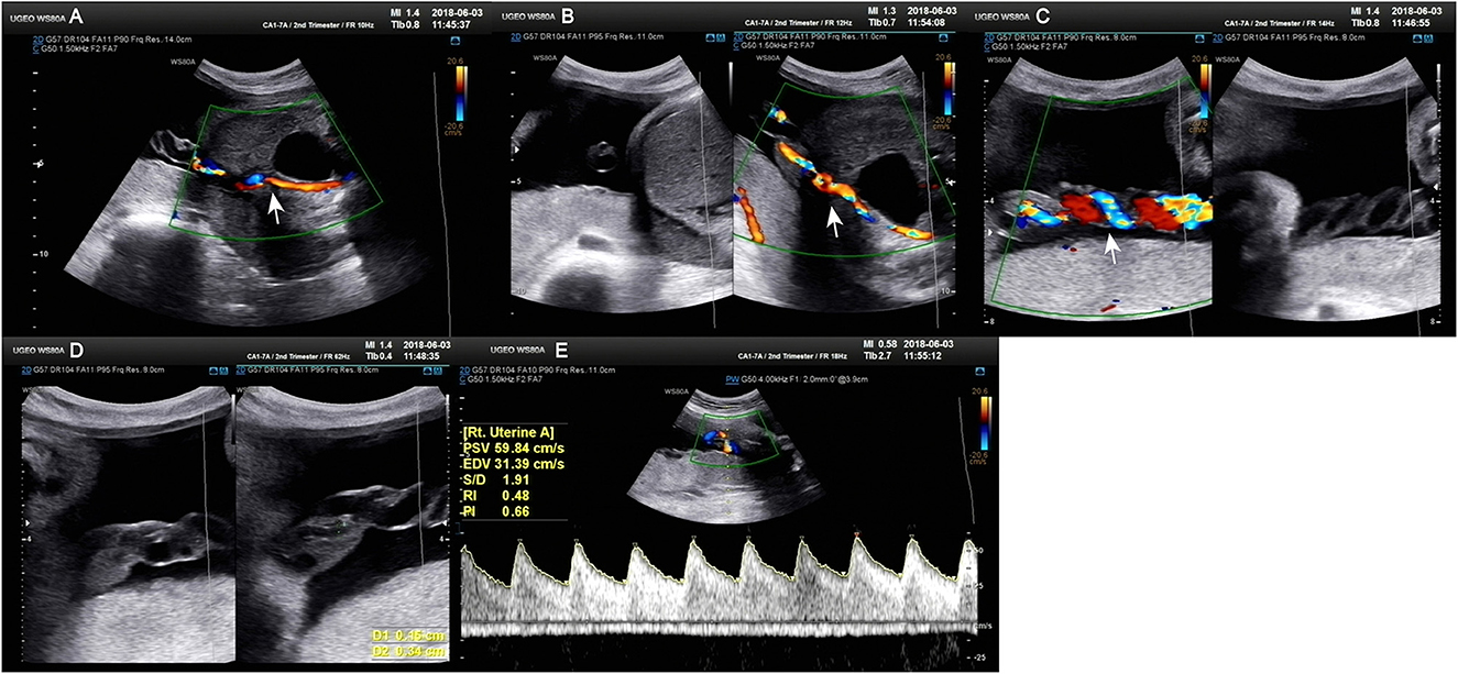
Frontiers | Umbilical artery thrombosis and maternal positive autoimmune  antibodies: two case reports and a literature review
Frontiers | Umbilical artery thrombosis and maternal positive autoimmune antibodies: two case reports and a literature review
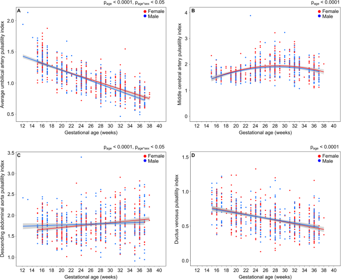
Fetal ultrasound scan of the third trimester

Sex differences in fetal Doppler parameters during gestation | Biology of  Sex Differences | Full Text
Sex differences in fetal Doppler parameters during gestation | Biology of Sex Differences | Full Text

Identifying the High-Risk Fetus in the Low-Risk Mother Using Fetal Doppler  Screening | Global Health: Science and Practice
Identifying the High-Risk Fetus in the Low-Risk Mother Using Fetal Doppler Screening | Global Health: Science and Practice

Ultrasound angiology reference standards of fetal cerebroplacental flow in  normal Egyptian gestation: statistical analysis of one thousand  observations | Egyptian Journal of Radiology and Nuclear Medicine | Full  Text
Ultrasound angiology reference standards of fetal cerebroplacental flow in normal Egyptian gestation: statistical analysis of one thousand observations | Egyptian Journal of Radiology and Nuclear Medicine | Full Text
Pregnancy Week 34
Pregnancy Week 34

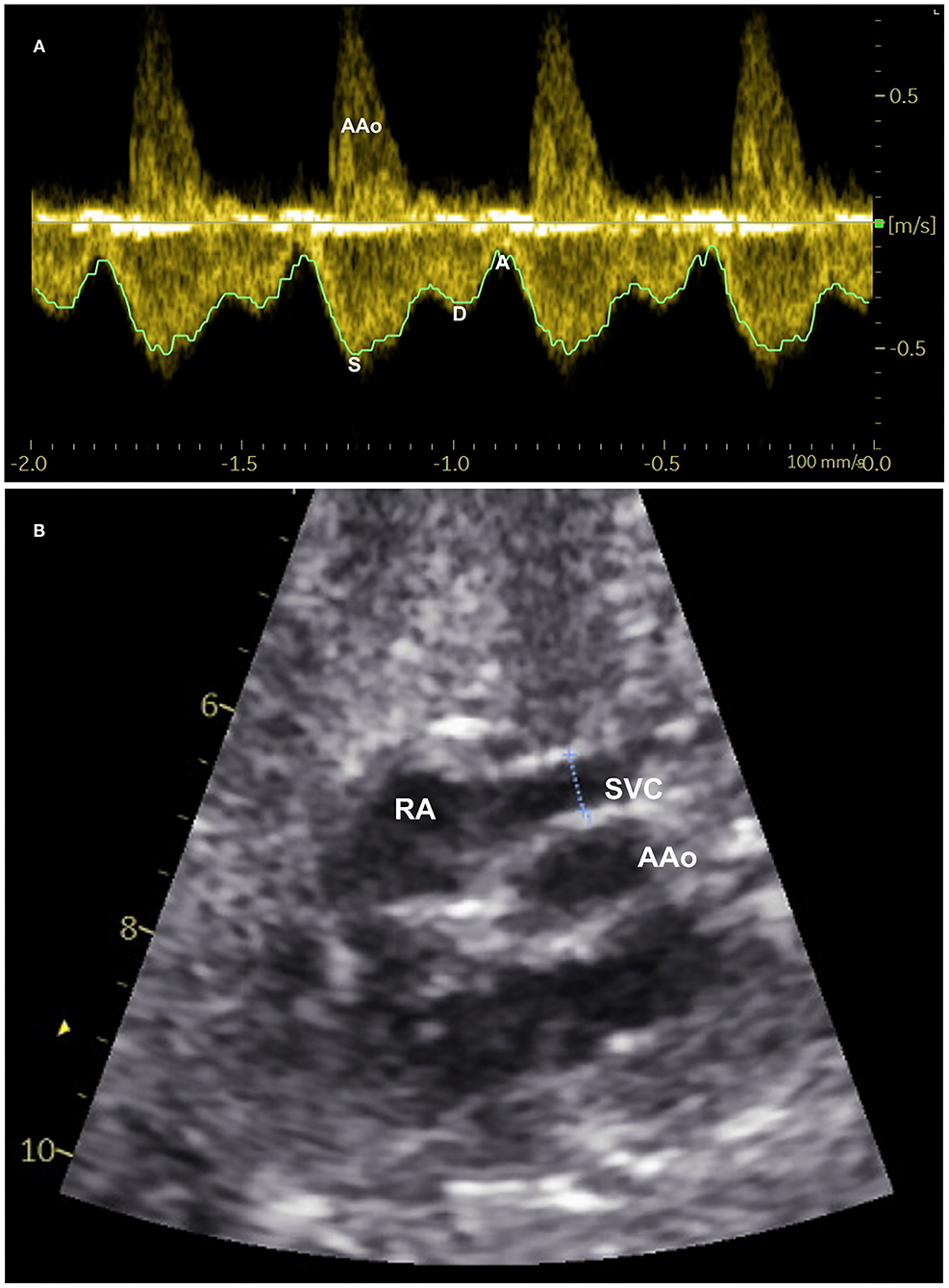
Frontiers | Fetal Superior Vena Cava Blood Flow and Its Fraction of Cardiac  Output: A Longitudinal Ultrasound Study in the Second Half of Pregnancy
Frontiers | Fetal Superior Vena Cava Blood Flow and Its Fraction of Cardiac Output: A Longitudinal Ultrasound Study in the Second Half of Pregnancy

shows the MCA Doppler indices in normal pregnancy at 34 weeks... | Download  Scientific Diagram
shows the MCA Doppler indices in normal pregnancy at 34 weeks... | Download Scientific Diagram

Color Doppler ultrasound and fetal brain magnetic resonance imaging... |  Download Scientific Diagram
Color Doppler ultrasound and fetal brain magnetic resonance imaging... | Download Scientific Diagram

Intrauterine Growth Restriction (IUGR) Imaging: Practice Essentials,  Ultrasonography
Intrauterine Growth Restriction (IUGR) Imaging: Practice Essentials, Ultrasonography

Palash - A Centre For Fetal Medicine - Growth and doppler study; This scan  is typically performed during the eighth month , around 32-34 weeks of  pregnancy. The purpose of this scan
Palash - A Centre For Fetal Medicine - Growth and doppler study; This scan is typically performed during the eighth month , around 32-34 weeks of pregnancy. The purpose of this scan

Antioxidants | Free Full-Text | Oxidative Stress in a Mother Consuming  Alcohol during Pregnancy and in Her Newborn: A Case Report
Antioxidants | Free Full-Text | Oxidative Stress in a Mother Consuming Alcohol during Pregnancy and in Her Newborn: A Case Report

34 Week Ultrasound - Reveal 3D/4D Ultrasound Studio

Assessment of Fetal Compromise by Doppler Ultrasound Investigation of the  Fetal Circulation | Circulation
Assessment of Fetal Compromise by Doppler Ultrasound Investigation of the Fetal Circulation | Circulation
Third Trimester Ultrasound Pictures

shows the uterine artery Doppler indices in normal pregnancy at 34... |  Download Scientific Diagram
shows the uterine artery Doppler indices in normal pregnancy at 34... | Download Scientific Diagram

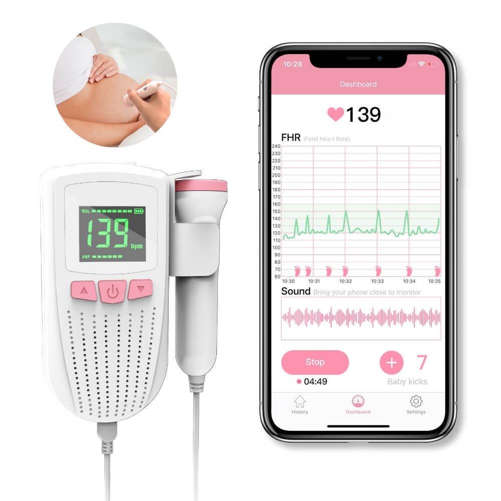
Fetal Doppler. Prenatal Baby Heart Beat Monitor. APP for Long-term  Tracking. Pocket FHR Detector. Highly sensitive Large Probe. Hear Baby  Sound. – Wellue
Fetal Doppler. Prenatal Baby Heart Beat Monitor. APP for Long-term Tracking. Pocket FHR Detector. Highly sensitive Large Probe. Hear Baby Sound. – Wellue

You are 34 Weeks and 4 Days Pregnant - FamilyEducation